किपा:Proteinviews-1tim.png
No higher resolution available.
Proteinviews-1tim.png (७५० × ३०० pixels, file size: १८२ KB, MIME type: image/png)
Summary
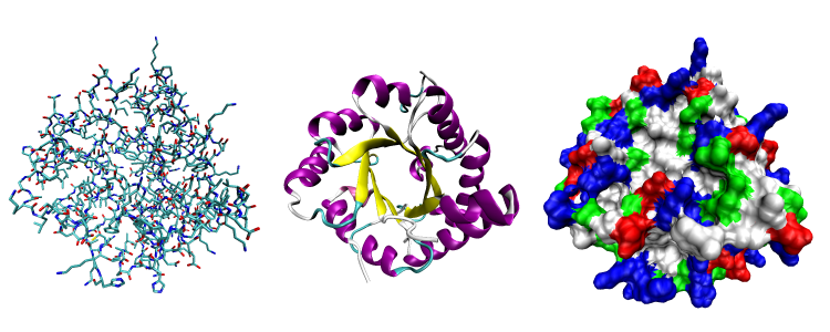
| Description | Three views of one monomer of the protein triose phosphate isomerase (PDB ID 1TIM). Left, an all-atom view colored by atom type; middle, a cartoon view colored by secondary structure; right, a solvent-accessible surface view colored by residue type (acidic residues red, basic residues blue, polar residues green, nonpolar residues white). |
| Date | |
| Source | Self created from PDB entry 1TIM using the freely available visualization and analysis package VMD |
| Author | Opabinia regalis |
| Permission (Reusing this file) |
GFDL |
Licensing
I, the copyright holder of this work, hereby publish it under the following license:

|
Permission is granted to copy, distribute and/or modify this document under the terms of the GNU Free Documentation License, Version 1.2 or any later version published by the Free Software Foundation; with no Invariant Sections, no Front-Cover Texts, and no Back-Cover Texts. A copy of the license is included in the section entitled GNU Free Documentation License. |
| This file is licensed under the Creative Commons Attribution-Share Alike 3.0 Unported license. | ||
| ||
| This licensing tag was added to this file as part of the GFDL licensing update. |
File history
Click on a date/time to view the file as it appeared at that time.
| Date/Time | Thumbnail | Dimensions | छ्य्लामि | Comment | |
|---|---|---|---|---|---|
| current | ०३:१९, १९ अगस्ट २००६ | ७५० × ३०० (१८२ KB) | Opabinia regalis | {{Information |Description=Three views of one monomer of the protein triose phosphate isomerase (PDB ID 1TIM). Left, an all-atom view colored by atom type; middle, a cartoon view colored by secondary structure; right, a solvent-accessible surface view col |
File usage
The following page uses this file:
Global file usage
The following other wikis use this file:
- Usage on ar.wikipedia.org
- Usage on ba.wikipedia.org
- Usage on be.wikipedia.org
- Usage on bs.wikipedia.org
- Usage on da.wikipedia.org
- Usage on en.wikipedia.org
- Usage on fr.wikipedia.org
- Usage on hy.wikipedia.org
- Usage on id.wikipedia.org
- Usage on it.wikiquote.org
- Usage on ja.wikipedia.org
- Usage on mn.wikipedia.org
- Usage on pl.wikipedia.org
- Usage on pl.wiktionary.org
- Usage on pt.wikipedia.org
- Usage on ru.wikipedia.org
- Usage on ru.wikibooks.org
- Usage on ru.wikiversity.org
- Usage on sh.wikipedia.org
- Usage on sl.wikipedia.org
- Usage on sr.wikipedia.org
- Usage on tr.wikipedia.org
- Usage on uk.wikipedia.org
- Usage on uz.wikipedia.org
- Usage on vi.wikipedia.org
- Usage on zh-classical.wikipedia.org
- Usage on zh.wikipedia.org